

15 May How Brain MRI Can Provide Insight Into Cognitive Disorders
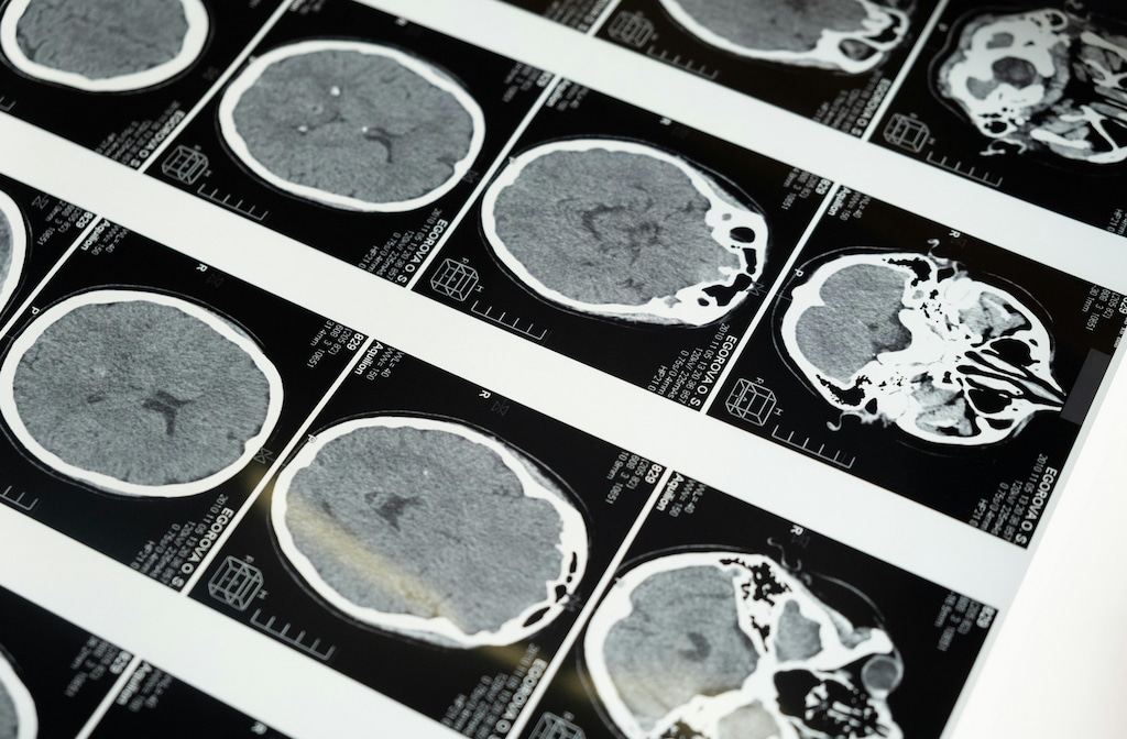
A brain MRI creates detailed images of brain tissue, blood vessels, and fluid spaces. Doctors use it to study structure, and they compare patterns across different brain regions. Since cognitive disorders affect memory, language, and behavior in different ways, MRI findings help narrow the cause. The scan does not diagnose every condition alone, but it adds clear data to the clinical picture.
Identifying Blood Vessel Damage
Small vessel disease often affects thinking speed, attention, and planning. A brain MRI shows white matter changes, and it also reveals old, small strokes. When blood flow problems injure deep brain tissue, these marks appear in specific patterns. Doctors review those patterns to separate vascular decline from other causes of memory loss.
Detecting Atrophy
Atrophy means loss of brain volume. MRI measures this change, and it tracks where tissue loss appears first. When shrinkage affects the hippocampus early, memory disorders move higher on the list. Doctors also assess whether the pattern looks focal or widespread.
Some disorders produce loss in the frontal or temporal lobes. MRI shows those regions clearly, but subtle changes may still need expert review. Since different syndromes damage different networks, the location of atrophy matters.
A careful reading helps connect scan findings with speech, judgment, or behavior changes. These findings, while invaluable, must always be interpreted in the context of the patient’s clinical presentation, as imaging alone cannot provide a definitive diagnosis, and a multidisciplinary approach often yields the most accurate conclusions.
Mapping Brain Activity
Structural MRI shows anatomy, while functional MRI tracks activity linked to blood flow. During testing, the scan records changing signals, and specialists map which regions engage during tasks. If language or memory networks respond in unusual ways, clinicians note that pattern. This method adds context when symptoms do not match a simple structural finding.
Functional imaging does not replace routine MRI. It adds another layer, and it helps describe how brain systems work together. When a team plans surgery or studies complex symptoms, this mapping may support safer decisions. In research and selected clinical cases, it offers a broader view of brain function.
Monitoring Chronic Conditions
Cognitive symptoms may change over time. Follow-up MRI tracks new lesions, and it measures whether atrophy progresses. When doctors compare scans across months or years, they look for stable findings or new damage. That timeline helps them judge whether a disorder remains steady or advances.
MRI also helps monitor conditions linked with ongoing cognitive change, such as:
- Multiple sclerosis
- Normal pressure hydrocephalus
- Prior stroke
- Traumatic brain injury
These disorders affect the brain in different ways, but imaging gives a repeatable record. If symptoms shift after treatment changes or new medical events, a fresh scan may clarify why. Doctors then compare image findings with testing results and daily function.
Schedule a Brain MRI
A brain MRI offers detailed information about structure, blood vessel injury, tissue loss, and network function. It does not answer every question, but it gives doctors a strong base for further evaluation. If you have memory changes, language problems, or ongoing confusion, speak with your physician about next steps. Schedule a brain MRI if your care team recommends imaging and needs clearer data.
Sorry, the comment form is closed at this time.